This is an old revision of the document!
Table of Contents
Thermal Injuries
Introduction
Skin is comprised of three anatomical layers: the epidermis, dermis, and subcutaneous tissue with each layer losing their function following a burn injury (Llyod et al., 2012)v. The epidermis layer acts as a barrier to bacteria and moisture loss, while the dermis provides elasticity and protection from trauma. The dermis also contains blood vessels that supply all the skin layers. When skin gets damaged, epidermal cells regenerate cells from deep within the dermal appendages, causing scarring (Llyod et al., 2012).
The International Society of Burn Injuries defines a burn as an injury sustained to the skin or any other organic tissue that is caused mainly by thermal or other acute trauma (Anandani, 2010). A burn occurs when some of all of the cells in the skin or other tissues get destroyed by contact with hot liquids (scalds), hot solids (contact burns) or through flames (flame burns). Other ways to receive burn injuries include radioactivity, friction, radiation, and contact with chemicals or electricity.The degree of tissue damage that is sustained through an injury depends on various factors (Anandani, 2010). These factors include the duration of contact with the source of the heat, the site of contact, the amount of heat energy, and sometimes also the agent itself which causes the burn.
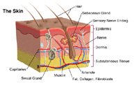 Figure 1- Anatomy of the skin. Retrieved from:http://www.hopkinsmedicine.org/healthlibrary/conditions/dermatology/burns_85,P01146/
Epidemiology
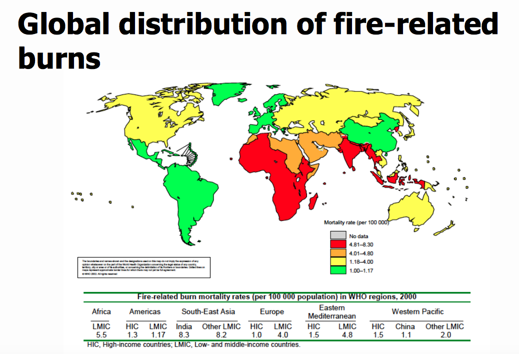
Globally, burns are a serious public health problem. Burns are the fourth most common type of trauma worldwide, following traffic accidents, falls, and interpersonal violence. An estimated 265 000 deaths occur each year from fires alone, with more deaths from scalds, electrical burns, and other forms of burns, for which global data are not available. Incidence varies by geographic location, socio-economic status, ethnic group, age and sex. Over 96% of fatal fire-related burns occur in low- and middle-income countries. In addition to those who die, millions more are left with lifelong disabilities and disfigurements, often with resulting stigma and rejection. High-income countries have made considerable progress in lowering rates of burn deaths, through combination of proven prevention strategies and through improvements in the care of burn victims. According to the World Health Organization, the highest mortality rates are observed in low and middle income countries with most cases seen in Southeast Asia. Mortality rate among low-income countries is 11 times higher than in high-income countries. For example: In India, over 1 000 000 people are moderately or severely burnt every year. Children under 5 and the elderly have the highest burn mortality worldwide.
Risk Factors
Factors that influence the incidence of burns injuries include age, sex, the home environment and economic status. Poverty is one of the main demographic factors associated with high risk of burns. It has been reported that children of low-income families are reported to have more than eight times the risk of sustaining burns compared to those children from higher income families (Edelman,2007). Furthermore, those children from the most deprived social class have burn-related death rates that are 25 times greater than children from affluent social classes (Edelman,2007). Limited education, illiteracy, family patterns and the type of residence are other risk factors associated with burn injuries. Housing locations in high-density areas such as slums have been reported as significant risk factors for childhood burns in countries such as Bangladesh and Pakistan (Daisy et al.,2007).
Pathophysiology
The body responds to a burn injury in two ways: local response and systemic response. Locally, there is tissue damage and systemically, organ systems are affected as a result of the burn (DeSanti, 2005).
Local Response: Three Zones of a Burn
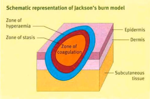
There are 3 zones of a burn, as described by Jackson’s Thermal Wound Theory.
- Zone of Coagulation: This is the most central location of the burn, where the damage is maximum and irreversible. This occurs due to coagulation of constituent proteins.
- Zone of Stasis: In this area there is decreased tissue perfusion (flow of blood). The tissue here can potentially be salvaged if it is not subject to additional damage such as an infection.
- Zone of Hyperemia: This is the peripheral area of the burn where there is an increased flow of blood and decreased cell injury. This area generally recovers unless there is additional damage such as an infection. (Hettiaratchy 2004).
Three Zones of a Burn
Systemic Responses
Cardiovascular Response
There are 2 stages of cardiovascular response following a burn injury: acute/resuscitative phase, which lasts about 48 hours and a hypermetabolic phase. During the acute phase, there is decreased blood flow to tissues and organs due to the direct effect of heat. In this phase, there is an increase in capillary permeability, which further induces fluid and protein loss. During the hypermetabolic phase, there in an increase in internal temperatures and blood flow to tissues and organs. In this phase, there is formation of an edema as well as increased water permeability. This can lead to cardiac instability, hypotension and decrease in myocardial contractility. It may dispose an individual to myocardial infarction (Cakir 2004) (Hettiaratchy 2004).
Pulmonary Response
Within the first couple hours of a burn injury, lung inflammation and lipid peroxidation occurs, which increases oxygen radicals. This response persists for a minimum of 5 days following the burn. Mortality can occur among burn victims if there are respiratory difficulties from inhaling smoke. Inhalation injury after burn trauma includes bronchial obstruction, airway resistance and altered capillary permeability. In severe burns, adult respiratory distress syndrome can occur (Hiettiaratchy 2004).
Renal Response
The renal system of an individual responds by decreasing renal blood flow and glomerular filtration rate after a burn injury. Levels of stress hormones, such as catecholamine, angiotensin, aldosterone and vasopressin increase. These renal responses can result in acute renal failure, which can further result in mortality (Cakir 2004).
Gastrointestinal Response
The renal system of an individual responds by decreasing renal blood flow and glomerular filtration rate after a burn injury. Levels of stress hormones, such as catecholamine, angiotensin, aldosterone and vasopressin increase. These renal responses can result in acute renal failure, which can further result in mortality (Cakir 2004).
Immune Response
Thermal injury results in an immunosuppressed state. Following a burn, the body releases pro-inflammatory factors but there is a depression in the lines of defense. When the skin layers are damages, there can be microbial invasion and growth in the damaged skin. Neutrophil accumulation increases in tissues, phagocytic activity is inhibited, macrophage hyperactivity increases and T-cells cannot function properly. This results in an increase in reactive nitrogen intermediated which cause immune dysfunction. Due to this, there is an increased susceptibility to sepsis, the presence of bacteria and toxins in tissues, and this can result in multiple organ failure. Burn toxins and oxygen radicals are also produced, which further lead to peroxidation. Comprised immune function is a leading cause of morbidity and mortality in burn patients (Cakir, 2004) (Hettiaratchy, 2004).
Types of Burns
Thermal Burns
Thermal burns occur due to heat sources coming in contact with the skin and raising the temperature. They can be caused by exposure to hot charring liquids, flames, steam, hot metals etc. This can result in tissue cell death as well as charring.
Electrical Burns
Electrical burns occur through electrocutions, when an electric current travels through the body.
Chemical Burns
Chemical burns occur when the skin or eyes come in contact with chemical products that are corrosive agents. Alkalis, acids, detergents or solvents can cause this type of burn.
Radiation Burns
Radiation burns occur when the skin is exposed to sources of radiation, such as UV from the sun or X-rays, for a long time (John Hopkins Medicine 2007).
Classifications of Burns
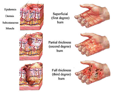
First Degree (Superficial)
First-degree burns directly affect the outer layer of the skin (epidermis). The burn site appears to be dry and red. An example of a first-degree burn is a sunburn.
Second Degree (Partial Thickness)
Second-degree burns affect the epidermis and dermis layers of the skin. The burn site appears to be red, blistered and swollen. An example of a first-degree burn is a burn from scalding hot water.
Third Degree (Full Thickness)
Third-degree burns fully penetrate the epidermis and dermis layers. The burn site appears to be white or charred but there are no sensations of pain since the nerves are destroyed in these areas. An example of a third-degree burn is a flame burn from a fire.
Fourth Degree
Fourth-degree burns occurs when bones, muscles and tendons are burned in addition to the epidermis and dermis layers. They can occur from prolonged exposure to flame or an electrical injury from high voltage. (John Hopkins Medicine 2007).
Degrees of a Burn
Assessment and Diagnosis
Management
Prevention
Providing adequate burn care in the Western world costs around $1000 US dollar per patient per day (Atiyeh et al.,2009).. This is not an ideal situation for those in developing countries and emphasizes the fact that prevention is a primary means to reduce burn-related death and disabilities. Barriers to prevention in developing countries also include having limited resources, limited knowledge in regards to first-aid treatment and inaccessibility to modern medicine (Atiyeh et al.,2009).. The primary target group of prevention efforts should be children from a lower socioeconomic status. This group is the largest and most vulnerable group for burn injuries. It is important to recognize that many of the risk factors associated with burn injuries cannot be modified easily or quickly and thus a multifaceted solution is needed for burns prevention. A multifaced solution requires the help of health workers to promote burn prevention along with programs implemented to help fight issues such as educational deficits, poverty, overcrowding, and poor housing (Atiyeh et al.,2009).
Atiyeh et al. (2009) highlight three main strategies that aim to reduce harm from injuries which include education, product design and environmental change, and legislation and regulation. In the strategies figure, strategies to reduce harm from injuries emphasize an active and passive approach. The educational strategy is an active approach focused on the individual (host) to help them avoid injury by modifying their environment to reduce the likelihood of injury. By knowing the environmental risks, it is more likely that preventative behavior will happen in an individual. Passive injury prevention involves product and environment modification. Product modification can be influenced by education the public to ask for safer products, and they can create pressure on authorities to produce prevention legislations (Atiyeh et al.,2009).
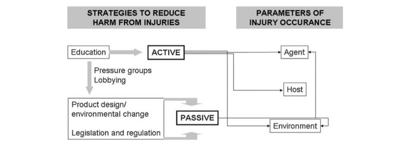 Fig. 1 – Strategies to reduce harm from injury. (Atiyeh et al., 2009)
PowerPoint Presentation
References
Alharbi, Z., Piatkowski, A., Dembinski, R., Reckort, S., Grieb, G., Kauczok, J., & Pallua, N. (2012). Treatment of burns in the first 24 hours: simple and practical guide by answering 10 questions in a step-by-step form. World Journal of Emergency Surgery, 7(1), 13.
Atiyeh, B. S., Costagliola, M., & Hayek, S. N. (2009). Burn prevention mechanisms and outcomes: pitfalls, failures and successes. Burns, 35(2), 181-193.
Anandani, J.H. (2010). Impact of thermal injury on hematological and biochemical parameters in burnt patients. Bioscience Biotechnology Research Communications: 3(1): 97-100.
“Burns.” John Hopkins Medicine. Retrieved January 22, 2017. http://www.hopkinsmedicine.org/healthlibrary/conditions/dermatology/burns_85,P01146/
Cakir, Baris and Berrak Yegen (2004). Systemic Responses to Burn Injury. Turk J Med Sci: 34: 215-226. http://journals.tubitak.gov.tr/medical/issues/sag-04-34-4/sag-34-4-1-0405-1.pdf
Daisy, S., Mostaque, A. K., Bari, S., Khan, A. R., Karim, S., & Quamruzzaman, Q. (2001). Socioeconomic and cultural influence in the causation of burns in the urban children of Bangladesh. Journal of Burn Care & Research, 22(4), 269-273.
DeSanti, Leslie (2005). Pathophysiology and Current Management of Burn Injury. Advances in Skin & Wound Care: 18(6):323-332.
Edelman, L. S. (2007). Social and economic factors associated with the risk of burn injury. Burns, 33(8), 958-965.
Hettiaratchy, Shehan and Peter Dziewulski (2004). Pathophysiology and Types of Burns. BMJ: 328(7453):1427-1429. https://www.ncbi.nlm.nih.gov/pmc/articles/PMC421790/
Lloyd, E.C.O., Rodgers, B.C., Michener, M., & Williams, M.S. (2012). Outpatient burns: Prevention and care. American Family Physician, 85(1), 25-32.
Peck, M. D. (2011). Epidemiology of burns throughout the world. Part I: Distribution and risk factors. Burns, 37(7), 1087-1100.
Peden M., McGee K., Sharma G. (2002). The injury chart book: a graphical overview of the global burden of injuries. Geneva, World Health Organization.
WHO. (2016). Fact Sheet on Burns. Violence and Injury Prevention, World Health Organization. Retrieved January 20, 2017 from http://www.who.int/mediacentre/factsheets/fs365/en/
WHO. (2007). Management of burns. Retrieved from http://www.who.int/surgery/publications/Burns_management.pdf
