This is an old revision of the document!
Table of Contents
What is Yellow Fever?
Symptoms and Prognosis
Yellow fever has an incubation rate of 3-6 days typically and will usually present itself with mild or no symptoms at all. (CDC, 2015) Infection begins with the “acute” phase where symptoms can include fever, chills, muscle pain, shivers, headache, loss of appetite, nausea and vomiting. Roughly 85% of people infected with the virus improve in a matter of several days, however the remaining affected will go into a more severe phase, usually a day after the onset of the infection. Symptoms for this include high fever, rapid jaundice, vomiting and abdominal pain. Mortality occurs in half of these patients within 10-14 days from the onset. (WHO, 2014).
Transmission
The main vector of transmission of Yellow Fever is the Aedes aegypti mosquito but is not limited to other types of mosquitoes such as Aedes albopictus (also known as the tiger mosquito). (Fontenille et al., 1997) Female mosquitoes obtain the virus by drinking the blood of primates or humans infected with the virus. The virus can then be transmitted the same way, through a mosquito bite. (CDC, 2015) There are three transmission stages: sylvatic, intermediate and urban. The first stage known as sylvatic is based in tropical rainforests where monkeys are infected as a result of vector mosquitos. These mosquitos may infect humans entering the forest. The second stage is intermediate and is based in humid or semi-arid areas of Africa where mosquitos can infect both humans and primates. This stage is the most common in Africa and has the potential to grow into an occasional epidemic. The third stage is known as urban and occurs in highly populated areas where the potential for an epidemic to occur is the greatest. Usually in these areas there are many unvaccinated people and vector mosquitoes. (WHO, 2014)
Epidemiology
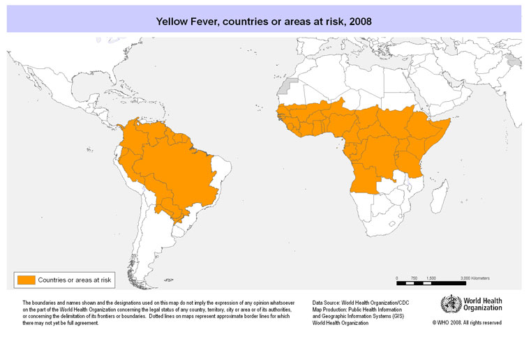
There are roughly 200,000 cases of yellow fever every year, of which 90% occur in Africa. It leads to 30,000 deaths annually, with the largest incidence rate being in Nigeria. (Monath, 2001). The virus persists in tropical and subtropical areas of Africa and South America but in recent years has been very prevalent in sub-Saharan regions of Africa and South America. Even with the presence of the vector mosquito in some regions and continents such as Asia and Australia, the virus has not affected individuals living there. The high prevalence of the virus in Africa is reported to be directly correlated with the high population of mosquitos living in close proximity to unvaccinated people. Due to the higher number of vaccinated people in South America, the transmission rate is much lower. Generally, obtaining reputable and accurate information regarding the transmission rate of the virus is difficult due to unreported cases, limited surveillance and recording techniques. (Barnett, 2007)
Figure 1: This image from the Public Health Agency of Canada shows the areas that are currently affected by the Yellow Fever Virus. Taken from: http://www.phac-aspc.gc.ca/publicat/ccdr-rmtc/10vol36/acs-11/index-eng.php
What virus causes Yellow fever?
Classification
Yellow fever is caused by the Yellow Fever Virus (YFV), which is part of the flavivirus genus. This group of disease-causing viruses consist of approximately 70 human or veterinary pathogens and are mostly spread through contact with a mosquito or a tick (Stock et al., 2013). Other prominent viruses that are part of the flavivirus genus include the West Nile virus, Dengue virus, Tick-borne Encephalitis virus and also the more recent Zika virus. Originating in Africa, the first case of flaviviruses in America was in the 17th century. Flaviviruses are all similar in size, with diameters ranging from 40-65 nm (Stock et al., 2013). This family of viruses also have relatively the same genome length which is consistently between 9500 and 12500 nucleotides. Full-length genome sequencing of all lineages of the virus examined over the span of decades showed little nucleotide variability, suggesting that the virus has a slow evolution rate and is genetically stable (Stock et al., 2013).
Genomic Sequence/ Structure
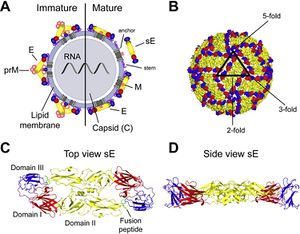
The Yellow Fever Virus (YFV) is a single-stranded RNA genome consisting of 11kb of nucleotides that code for polyproteins (Volk et al., 2009). The virus consists of a virion that includes three structural proteins and seven non-structural proteins. The structural proteins along with the RNA genome form something called the flavivirus virion, which is the complete and infective form of the virus. In terms of the YFV, the virion is 50 nm in diameter and is composed of the E protein, M protein and a capsid protein essentially acting as an envelope to protect the RNA of the virus (Patkar et al., 2007). The largest, most significant structural protein is the envelope (E) protein. This protein is the main immunogen and is essential during receptor binding and membrane fusion. Once binding to the host cell occurs, the capsid protein is released into the cytoplasm of the infected cell (Patkar et al., 2007). In contrast, the non-structural proteins are responsible for various enzymatic activities such as those of RNA Helicase and Protease but are not expressed in the flavivirus virion.
Figure 2:Genomic sequence showing different structural and non-structural components of virus. (Rice et al., 1985).
Yellow Fever Viruses are positive-sense, single-stranded and are generally in the shape of icosahedron, meaning they have 20 sides. The fact that the RNA strand is positive-sense provides major significant advantages to the virus during cell entry and replication. By being positive-sense, the viral RNA has the ability to be directly translated into the viral protein since the RNA can be considered the mRNA as well (Rice et al., 1985). Furthermore, YFV, unlike other mosquito-borne diseases is viscerotropic, as it preferentially affects the main organs in the internal cavity of the body such as the kidneys (Rice et al., 1985).
Figure 3: Top view and side view of Yellow Fever Virus, specifically showing immature and mature structures and also highlighting the three domains of the virus structure. Taken from: http://www.thenextgenscientist.com/page/3/
How does Virus Cause Yellow Fever?
Immune System Response
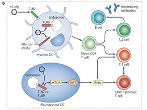
Research has shown that the immune system responses to combat yellow fever can be observed after administering the yellow fever vaccine. Once an individual is vaccinated, researchers are able to analyze the types of immune cells that interact and limit the spread of yellow fever infection (Pulendran, 2009). This is possible either through using a genetic screen that identifies genes which are expressed by immune cells or using flow cytometry to determine specific proteins that are expressed by immune cells. Using these methods, the immune cells that have been identified are dendritic cells (DC), a variety of T cells and B cells (Querec et al., 2009; Gaucher et al., 2008). DCs are important for the immune response against yellow fever as they are the main activators of other adaptive immune cells that directly kill yellow fever infected cells. Research has shown that specific receptors present on the DC called toll-like receptors are able to bind to yellow fever antigens. Upon binding, the DC can then present the yellow fever antigen to naïve CD4+ T cells resulting in the activation of the apoptotic CD8+ T cells as well as neutralizing antibodies that can directly interfere with yellow fever activity in the blood. Please see Figure below for details. Ultimately, immune system recognition of yellow fever results in a well rounded and robust response that involves effective communication between multiple cell types (Pulendran, 2009).
Figure 4: The image represents a diagram showing the different parts involved in the immune response to the Yellow Fever virus. Taken from: http://www.nature.com/nri/journal/v9/n10/full/nri2629.html
Immune System Evasion
Current procedures
Yellow Fever has no cure (Bukowski, 2015). Since there is no cure, managing the disease becomes essential. Once infected, supportive care is critical and, if conditions worsen, the patients are treated in an intensive care setting (Bukowski, 2015). The infection causes a decline in blood pressure, for which the patients are given vasoactive medication which works as a vasoconstrictor, thus increasing the patients' blood pressure (Bukowski, 2015). Symptoms such as excessive bleeding and vomiting will cause a huge fluid imbalance in the body. However, administering intravascular fluid will keep the body hydrated and also keep the blood pressure high (Bukowski, 2015). Also, ventilator management is used to support patients' breathing, as these machines give oxygen to the lungs (Bukowski, 2015). Furthermore, you would have to administer the treatment of disseminated intravascular coagulation (DIC), hemorrhage, secondary infections, and renal and hepatic dysfunction. For actively bleeding patients, the administration of fresh frozen plasma is recommended to maintain a prothrombin time of 25-30 seconds (Bukowski, 2015). In patients with DIC, heparin has been a recommended for treatment (Bukowski, 2015).
Vaccines
There are two different sub strains derived from the YF-17D, YF-17D-204 and YF-204, which are used for the production of yellow fever (Roukens & Visser, 2008). It is grown in an embryonated chicken egg, yielding 100-300 vaccines doses per egg and it should be used immediately after produced (Roukens & Visser, 2008). This vaccination can prevent yellow fever and the current vaccine for yellow fever confers 95% lifelong immunity in patients (Roukens & Visser, 2008). Based on the severity of Yellow fever in certain areas, it is recommended to have routine immunization. Currently, world health organization strives for an 80% coverage for the vaccine to achieve immunity for many people to prevent an outbreak (Roukens & Visser, 2008). For travelers from non-endemic areas to endemic areas it is crucial to take this vaccine.
Safety
This vaccine has been administered to over 500 million people worldwide, and has been regarded as a safe drug (Roukens & Visser, 2008). The adverse effects usually occur within 2-6 days after vaccination and include low grade fever, headache, myalgia headache, and redness at the site of the injection (Roukens & Visser, 2008). Only 10-20 % of these symptoms are reported. This vaccination is advised for adults and children over the age of 9 months and under 65 years (Roukens & Visser, 2008). Furthermore, anaphylaxis can occur after vaccination with a risk of 0.8 in 100000 doses. Obtaining history on patients for hypersensitivity to eggs or egg products can reduce the risk of anaphylaxis (Roukens & Visser, 2008). The reason for this is that because this vaccine is grown in an embryonate chicken egg. To test hypersensitivity of patients with egg allergies, they give 0.1ml to patients under strict supervision (Roukens & Visser, 2008). Then a controlled saline dose is given to the other arm and both sites are checked after 30 minutes. If the diameter of the cutaneous wheal of the yellow fever virus is less than twice the control diameter, hypersensitivity to the vaccine is highly unlikely. If it exceeds the diameter two folds, the yellow fever vaccine will not be administered. Moreover, yellow fever vaccination in early pregnancy stages is safe (Roukens & Visser, 2008).
Future procedures?
Over the past 60 years the YF-17D vaccine has not changed. There are limitations on the amount of vaccinations that can be produced on a short notice. This a concern to the Global Alliance of Vaccines, which is supporting the stock pile of over 6 million doses of yellow fever vaccines (Roukens & Visser, 2008). According to the WHO, shortages in vaccine supply is soon to become a problem. In order to make it more economically friendly, they will reduce standard dosages fivefold from 0.5ml to 0.1ml (Roukens & Visser, 2008). Furthermore, injecting inactive yellow fever vaccines in people traveling to endemic areas may provide protection, as there has been trial with the inactive vaccine in mice, finding that all the mice were protected from the virus (Roukens & Visser, 2008).
Summary
References
1. Gaucher, D., Therrien, R., Kettaf, N., Angermann, B. R., Boucher, G., Filali-Mouhim, A., … & Akondy, R. (2008). Yellow fever vaccine induces integrated multilineage and polyfunctional immune responses. The Journal of experimental medicine, 205(13), 3119-3131.
2. Pulendran, B. (2009). Learning immunology from the yellow fever vaccine: innate immunity to systems vaccinology. Nature Reviews Immunology, 9(10), 741-747.
3. Querec, T. D., Akondy, R. S., Lee, E. K., Cao, W., Nakaya, H. I., Teuwen, D., … & Kennedy, K. (2009). Systems biology approach predicts immunogenicity of the yellow fever vaccine in humans. Nature immunology, 10(1), 116-125.
4. CDC (2015, August 13). Yellow Fever: Symptoms and Treatment. Retrieved from http://www.cdc.gov/yellowfever/symptoms/index.html
5. WHO (2014, March 1). Yellow Fever. Retrieved from http://www.who.int/mediacentre/factsheets/fs100/en/
6. CDC (2015, August 13). Yellow Fever: Transmission. Retrieved from http://www.cdc.gov/yellowfever/transmission
7. Fontenille, D., Diallo, M., Mondo, M., Ndiaye, M., & Thonnon, J. (1997). First evidence of natural vertical transmission of yellow fever virus in Aedes aegypti, its epidemic vector. Transactions of the Royal Society of Tropical Medicine and Hygiene, 91(5), 533-535.
8. Barnett, E. D. (2007). Yellow Fever: Epidemiology and Prevention. Clinical Infectious Diseases, 44(6), 850-856.
9. Monath, T. P. (2001). Yellow fever: An update. The Lancet Infectious Diseases, 1(1), 11-20.
10. Volk, D.E., May, F.J., Gandham, S.H., Anderson, A., Von Lindern, J.J., Beasley, D.W., Barrett, A.D., & Goreinstein, D.G. (2009). Structure of Yellow Fever Virus Envelope Protein Domain III. US National Library of Medicine, 394(1), 12-18.
11. Patkar, C.G., Jones, C.T., Chang, Y., Warrier, R., & Kuhn, R.J. (2007). Functional Requirements of the Yellow Fever Virus Capsid Protein. Journal of Virology, 81(12), 6471-6481.
12. Stock, N.K., Laraway, H., Faye, O., Diallo, M., Niedrig, M., & Sall, A.A. (2013). Biological and Phylogenetic Characteristics of Yellow Fever Virus Lineages from West Africa. Journal of Virology, 87(5), 2895-2907.
13. Rice, C.M., Lenches, E.M., Eddy, S.R., Shin, S.J., Sheets, R.L., & Strauss J.H. (1985) Nucleotide Sequence of Yellow Fever Virus: Implications for Flavivirus Gene Expression and Evolution. Science, 229(4715), 726-733
14. Busowski, M. T. (2015, June 26). Yellow Fever. Retrieved,from http://emedicine.medscape.com/article/232244-overview
15. Roukens, A. H., & Visser, L. G. (2008). Yellow fever vaccine: past, present and future. Expert opinion on biological therapy, 8(11), 1787-1795.


