Table of Contents
Global Burden of Disease: Coronary Artery Disease (CAD)
Coronary Artery Disease Powerpoint
Introduction
Global Burden of Disease
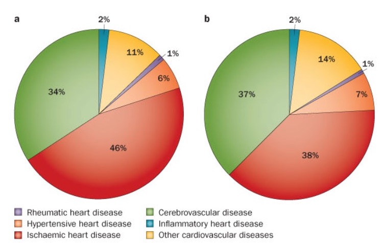
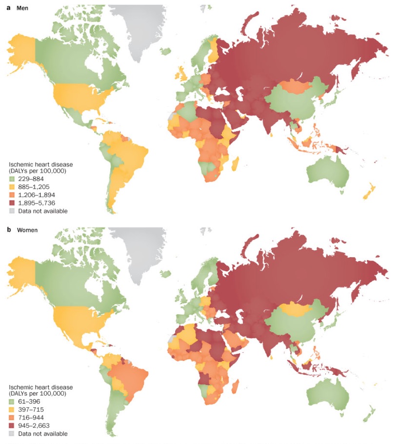
The global burden of disease measures the burden of a specific disease using the disability-adjusted-life-year (DALY) measurement. This measure combines the years of life lost due to premature mortality and the years of life lost due to time lived in states of less than full health. The global burden of disease is the highest for ischaemic heart disease or coronary artery disease (CAD). CAD is the leading cause of mortality burden, morbidity and loss of quality of life (Mathers and Loncar, 2006).
Coronary Artery Disease
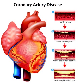
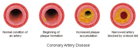
Coronary Artery disease (CAD), also known as Coronary Heart Disease (CHD) and Ischaemic heart disease is the largest contributor to cardiovascular diseases (Wong 2014). The diseases are characterized by: angina (chest pain), shortness of breath and myocardial infarction which may contribute to patient mortality. One of the main causes of CAD is atherosclerosis which is characterized by gradual thickening of the coronary artery walls. This occurs when plaque, composed of debris, fat and organic substances accumulates inside the arterial walls, leading to narrowing and hardening of the blood vessels.
Other risk factors include hypertension, cigarette smoking, diabetes mellitus or elevated glucose levels, increased cholesterol levels, and obesity (Wong, 2014). Specifically, a dangerous combination of poor nutrition and lack of physical exertion can lead to many of these risk factors. Current research suggests preventive measures are the most beneficial in reducing an individual's risk for developing CAD; this includes proper nutrition and doing regular exercise. Furthermore, additional treatment options include the use of medication such as beta blockers, nitrates, angiotensin converting enzyme (ACE) inhibitors, and antiplatelets. Surgery is a more invasive option for those afflicted with multiple blockages and the goal is to improve circulation of blood towards the heart.
Epidemology
Framingham Heart Study
CAD is a major cause of death and disability in developed countries, although mortality rates have been observed to be decreasing over the last decades in western countries (Sanchis-Gomar et al 2016). Despite the decline, CAD still causes approximately one-third of all deaths in people older than 35 years. The Framingham Heart, which is an ongoing population-based cardiovascular cohort study concludes the many risk factors that contribute to CAD. The study concluded that men over the age of 38 had 1.5 times greater risk of developing atrial fibrillation than women (Benjamin et al 1994).
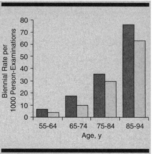
A follow up of the Framingham study based on 44 years of data and observation of the offspring of the original study members was conducted. Results indicated individuals aged over 40, the lifetime risk of developing CAD was 49% in men and 32% in women. The lifetime risk for 70 years old individuals was 35% in men and 24% in women. Lastly, for total coronary events, the incidence rose steeply with age, with women behind men by approximately 10 years.
Prevalence and Mortality Rate
The 2016 Heart Disease and Stroke Statistics update of the American Heart Association reported that 15.5 million people over 20 years of age in the USA have CAD, with the prevalence increasing with age for both sexes (Sanchis-Gomar et al, 2016). The results of the data collected on CAD and MI is illustrated in the graphs where the prevalence for men is observed to be much greater than women.
Furthermore, in a 2009 report based on data from National Health and Nutritional Survey (NHANES), myocardial infarction prevalence was observed in middle aged individual (35-45 years of age) during the 1988-1994 and 1999-2004 time periods. The results indicated a higher prevalence in men compared to women, but the trend declined for the former over time.
Global and Regional DALY
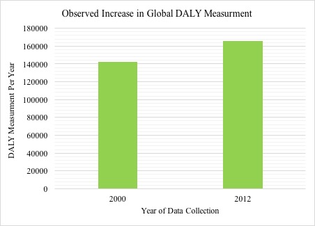
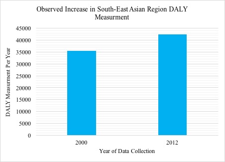
The World Health Organization released estimates of the global and regional burden of disease, which included 20 leading causes. The data was collected for 2000 and 2012, with a reported increase in DALY being observed both globally and in regions such as Southeast Asia, the Eastern Mediterranean and the western pacific region. The global rank of ischemic heart disease increased from being the third leading cause of death in 2000 to the first leading cause of death in 2012 (World Health Organization, n.d.) Although the global burden of coronary artery disease was dominant in western countries during the early 20th century, the burden now lies in specific Asian and middle-Eastern regions (Wong 2014). The global ischemic heart disease DALY data collected for both sexes indicates a high concentration in Central Asia, North Africa and West Europe.
Pathophysiology
Coronary Artery Anatomy
Arteries are shaped like hollow tubes and have muscular walls lined with a layer of cells known as the endothelium (Libby & Theroux, 2005). These walls are smooth and elastic and provide a physical barrier between the walls of the artery and the blood stream (Cleveland Clinic, 2017).
Atherosclerosis
One of the leading causes of CAD is atherosclerosis or the gradual thickening of the walls of the coronary arteries (Cleveland Clinic, 2017). Atherosclerosis occurs when plaque builds up inside of arterial walls, causing the narrowing and hardening of these blood vessels (Cleveland Clinic, 2017).
As individuals age, cellular debris begins to stick to the vessel walls. The amalgamation of debris, fat and other substances combine to create what is known as ‘plaque' (Libby & Theroux, 2005). Plaque can be composed of many things, but often includes cholesterol, fat substances, metabolic waste, calcium, and fibrin (a clotting compound found in the blood) (Libby & Theroux, 2005). The inside of the arteries develop plaques of different shapes and composition as a person ages (Libby & Theroux, 2005). Most plaques have a fibrous ‘cap’ covering a softer interior. If the cap either cracks or is torn, the soft inside is exposed, and platelets form a clot around the plaque (Cleveland Clinic, 2017). This can cause irritation in the endothelium, and even cause a disruption in function where the artery contracts inappropriately and thus narrows the artery even more (Cleveland Clinic, 2017).
Once broken off, the clot may disintegrate into smaller components, thus restoring normal blood flow patterns (Cleveland Clinic, 2017). However, what often happens is the thrombus (blood clot) begins to block blood supply to the heart muscle causing what is known as a coronary occlusion. This would be an acute cause of CAD, as it is the result of a sudden rupture of a plaque and formation of a thrombus (Cleveland Clinic, 2017). The alternate cause of CAD is of chronic origin- the coronary artery is simply narrowed over time and blood supply is slowly limited (Cleveland Clinic, 2017).
Atherosclerotic Lesions
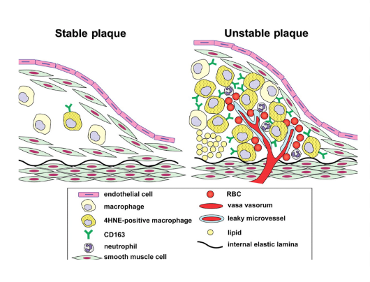
Beginning as smaller lesions, early atherosclerotic lesions can then rapidly enlarge into plaques in individuals with coronary risk factors (Fuster et al., 1992). Atherosclerotic lesions, or atheromata, are the asymmetric thickening of the intima (the innermost layer of the artery). Atheromata are composed of cells, connective tissue elements, lipids, and cellular debris (Fuster et al., 1992). The core of an atheroma is made up of immune cells such as T-cells and macrophages, which can produce inflammatory cytokines, coagulation factors, radicals, and proteases when activated. These factors can then, in turn, destabilize lesions and induce ischemic events (Fuster et al., 1992).
Ischemic Events & Plaque Disruption
An ischemic event occurs when there is a restriction or a reduction in blood supply to the tissues, causing a shortage in the materials needed for cellular metabolism in the cells (e.g. oxygen and glucose) (Fuster et al., 1992). In CAD, there are a variety of ways in which ischemic events can be triggered (Fuster et al., 1992).
Lipid Rich Plaques Small atherosclerotic plaques are often made up of a crescent-shaped mass of lipids, and are relatively small and soft due to both their high concentration of cholesterol and the presence of the chemical compound ester. If there is a change in shear force around the area of stenosis or a sudden change of intraluminal pressure, these small plaques may be disrupted and cause an ischemic event (Fuster et al., 1992).
Macrophages Responsible for the uptake and metabolism of lipids, macrophages are thought to play a role in the development of plaques. Macrophages may also play a role in plaque formation because they enhance the transport of cholesterol, and release a mitogenic factor that helps the proliferation of smooth-muscle cells. They play a role in plaque disruption as they release proteases that digest the extracellular matrix, which can then cause a plaque to be released (Fuster et al., 1992).
Vessel Wall Stress Plaques may also be disrupted due to alterations on or within plaques. Some plaques, known as fissured plaques, contain a fibrous cap that when under tensile stress, can rupture due to the lack of collagen support under the cap. Other forms of plaques may be distressed by vessel wall stress due to the disturbed blood flow pattern due to areas of stenosis (narrowing). This altered pressure and flow may cause plaque tearing. Finally, plaques may also be disrupted by the bending and twisting of the artery during contractions (Fuster et al., 1992).
Symptoms
CAD can present itself in a variety of ways, including:
Chest Pain (Angina) is typically described as tightness or pressure in your chest and normally occurs in the middle or left side. Typically induced by stress, the pain usually subsides within minutes of reducing exertion. Women tend to feel the pain in the neck, arm, or back instead of the chest (Mayo Clinic Staff, 2015).
Shortness of Breath is due to the obstruction or stenosis of arteries. In these cases, the heart is unable to pump blood to meet the needs of the body which can cause extreme fatigue (Mayo Clinic Staff, 2015).
Heart Attack or Myocardial Infarction (MI) can occur if the coronary artery is completely occluded. Symptoms of a heart attack include a crushing or squeezing pressure in the chest, pain in your shoulder or arm, shortness of breath, nausea, denial, or sweating. Women often experience different symptoms than men, such as pain in the neck or jaw. Heart attacks can also occur without any of the hallmark warning signs or symptoms, these are known as ‘silent Heart Attacks’ (Mayo Clinic Staff, 2015).
Diagnosing CAD
Typically, if a patient displays symptoms (shortness of breath, angina, etc) they would be referred to do one of these diagnostic tests to determine the presence of CAD. There are numerous diagnostic tests available for determining the presence of CAD, these include: electrocardiogram (ECG), echocardiogram, exercise stress test, and coronary angiogram.
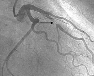
An ECG and echocardiogram are both non-invasive techniques and simply image the heart at rest (Mayo Clinic, n.d.). If a patient displays symptoms during exercise, an exercise stress test may be ordered (Mayo Clinic, n.d.). All of these tests can determine if a patient has CAD but typically more testing needs to be done to be absolutely sure. If either one of these tests is positive, a patient is referred for an Coronary Angiogram which is considered the gold standard for diagnosing CAD (Bogle, n.d.).
As you can tell, many of these diagnostic techniques require complex machines and the expertise of healthcare professionals to properly determine the presence or absence of disease. The combination of these two factors may be simply impractical for many living in impoverished communities around the world. Thus, without the proper tools to diagnose CAD, the current statistics for the global burden of this disease may actually severely underestimate its truly devastating impact.
Risk factors
Physiological Risk Factors
Physiologically, high cholesterol, high blood pressure, and high blood sugars (diabetes) have all been reported as key risk factors for CAD (Coronary Artery Disease Risk Factors, 2016). In addition, one’s risk for developing CAD increases greatly as the number of risk factors an individual possess increases (CADRF, 2016). Fortunately because of the advances in medicine, there are numerous pharmacological treatment options available for combating each and every single one of these aforementioned risks. Furthermore, there are also non-modifiable risk factors for which someone can do nothing about. These include age, sex, and ethnicity (CADRF, 2016).
Lifestyle Risk Factors
However, the truly problematic risk factors are the ones that are sociological in nature for which there are no easy fixes. For example, it has been shown that a combination of poor nutrition and lack of physical activity can lead to the development of obesity which can greatly increase one’s risk for developing CAD (Eckel & Krauss, 1998). It can seem obvious to state that if an obese individual wanted to lower their risk factors for CAD that they should lose weight. This simple statement is extremely difficult to implement as long term clinically significant weight loss is seldomly achieved (Wing & Phelan, 2005).
Healthcare Factors
To further complicate the problem, individuals in developing nations simply lack the access to affordable and reliable healthcare facilities to adequately treat their chronic conditions. In addition, healthcare systems around the world are reactionary in nature and tend to treat diseases only when the patient manifests symptoms rather than preventing the disease in the first place. This approach to healthcare is especially problematic for chronic diseases such as CAD for which there appears to be no immediate cure but rather the only plausible medical solution is to manage the current conditions to not allow any advancement in the disease. Unfortunately, this approach to disease is extremely expensive, and thus simply impractical for many living in developing nations. Thus without the proper medical management of CAD in addition to rising global life expectancies, especially in predominantly developing parts of the world, the global burden of CAD will undoubtedly increase for the foreseeable future (World Health Organization, n.d.). The most effective long term strategy for combating CAD should focus on preventing any and all risk factors associated with CAD from occurring in the first place.
Treatments
CAD can be treated through pharmacological options (medications), medical procedures and lifestyle modifications. According to data comparing CAD deaths from the 1980’s and 2000, it was estimated that about 47% of the decline in CAD deaths was attributed to treatments (Llyod-Jones et al., 2010).
Pharmacological Alternatives
There are numerous medications available that are used to lower blood pressure and prevent blood clots from forming which ultimately reduces your chances of mortality from CAD. These medications include: beta blockers, nitrates, angiotensin converting enzyme (ACE) inhibitors, and antiplatelets.
Beta blockers which include atenolol, metoprolol, nebivolol and bisoprolol are used to slow down the heartbeat and improve blood flow. Whereas, nitrates, often referred to as vasodilators are used to widen blood vessels and control angina symptoms. The ACE inhibitors, one of the most commonly used drugs are used in reducing the risks of a cardiovascular event, by lowering blood pressure. They work by blocking the conversion of angiotensin-I (AT-1) into angiotensin- II (AT-II) by blocking the main effects of AT-II which are; narrowing blood vessels and water and salt retention (Brugts et al.,2012). Another type of medicine commonly used is antiplatelet medicines include aspirin. Antiplatelet medicines are known to prevent the formation of blood clots. Aspirin, the most widely used antiplatelet agent is known to reduce the risks of vascular events by inhibiting platelet aggregation (Hennekens,Dyken & Fuster, 1997).
There are numerous medications available that are used to lower blood pressure and prevent blood clots from forming which ultimately reduces your chances of mortality from CAD. These medications include: beta blockers, nitrates, angiotensin converting enzyme (ACE) inhibitors, and antiplatelets. Beta blockers which include atenolol, metoprolol, nebivolol and bisoprolol are used to slow down the heartbeat and improve blood flow. Whereas, nitrates, often referred to as vasodilators are used to widen blood vessels and control angina symptoms. The ACE inhibitors, one of the most commonly used drugs are used in reducing the risks of a cardiovascular event, by lowering blood pressure. They work by blocking the conversion of angiotensin-I (AT-1) into angiotensin- II (AT-II) by blocking the main effects of AT-II which are; narrowing blood vessels and water and salt retention (Brugts et al.,2012). Another type of medicine commonly used is antiplatelet medicines include aspirin. Antiplatelet medicines are known to prevent the formation of blood clots. Aspirin, the most widely used antiplatelet agent is known to reduce the risks of vascular events by inhibiting platelet aggregation (Hennekens,Dyken & Fuster, 1997).
Medical Procedures
Patients with severe chest pain or severe obstruction of the coronary arteries require more invasive treatment options. Currently, there are numerous surgeries which can be performed to combat CAD. These procedures include: Coronary Artery Bypass Graft Surgery (CABG), and Percutaneous coronary intervention (PCI) (Llyod-Jones et al., 2010).
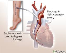
CABG the most common type of heart surgery, is performed to improve blood flow into the heart. As seen in figure 11, this surgery is performed by connecting a healthy artery or vein from another part of your body such as from the leg, chest, or arm to the blocked coronary artery. The healthy artery or vein bypasses the blocked or narrowed part of the artery, thus providing a new pathway for oxygen-rich blood to flow to the heart. During CABG, the breastbone is divided and the heart is stopped to send blood through a heart-lung machine. It is important to note that bypass surgery will not cure coronary artery disease, but it will improve heart function and reduce the risk of dying of heart disease (National Heart, Lung, and Blood Institute, 2016). After the surgery, it is important that the patient takes time to recover. During the recovery period, a patient may participate in a cardiac rehabilitation program under the direct supervision of medical professionals.
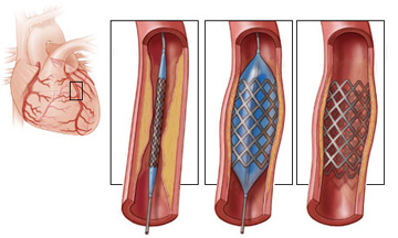
As seen in figure 12, PCI, which is also known as coronary angioplasty, is a nonsurgical procedure often used to open blocked or narrow coronary arteries. The procedure requires cardiac catheterization which involves the insertion of a hollow tube (catheter) with a small inflatable balloon at its tip covered with a stent. The inserted balloon catheter then compresses the plaque and opens up the blocked artery. The inserted stent then remains in the artery holding it open (National Heart, Lung, and Blood Institute, 2016).
Life Style Modifications
Lifestyle modifications are important for both preventing, and treating CAD. They include: proper nutrition, and regular physical activity.
Nutrition
In general, avoiding a lot of red meat, sugary foods and beverages, and trying to lower your consumption of saturated and trans fat are crucial for reducing your risk of CAD. Saturated fat and trans fat, are known to raise blood cholesterol levels and thus it's important to consume more sources of monounsaturated and polyunsaturated fats which are known to help lower blood cholesterol levels. For example, avocados, salmon & trout, nuts and seeds, olive, canola, peanut , tofu are all great sources of monounsaturated and polyunsaturated fats. In addition, limiting the amount of alcohol you consume and quitting smoking, are also essential in reducing your risks of CAD (National Heart, Lung, and Blood Institute, 2016).
Physical Activity
Regular physical activity has been shown to improve symptoms and reduce mortality in patients with CAD. According to a study conducted in 2001, patients in the training group experienced fewer cardiac events and had a lower number of hospital readmissions. The total mortality of patients with CAD was found to be reduced by about 27% and 31%, as a result of the exercise training (Erbs, Linke & Hambrecht, 2006), (Jolliffe, Rees, Taylor, Thompson, Oldridge,& Ebrahim, 2001).
Future Global Burden
The global burden of coronary artery disease (CAD) is expected to increase in the future. This is especially true in developing countries due to a large population size, low education level, poor lifestyle choices and poor healthcare facilities. According to the Global Burden of Disease Study, developing countries contributed 3.5 million of the 6.2 million global deaths from CAD in 1990 (Murray and Lopez, 1996). This study estimates that the developing countries will account for 7.8 million of the 11.1 million deaths due to CAD by 2020 (Murray and Lopez, 1996). This study provides evidence for an increased economic burden of CAD from only developing countries and all the countries of the world.
References
Benjamin, E. J., Levy, D., Vaziri, S. M., D'Agostino, R. B., Belanger, A. J., & Wolf, P. A. (1994). Independent risk factors for atrial fibrillation in a population-based cohort: the Framingham Heart Study. Jama, 271(11), 840-844.
Bogle, Richard. (n.d.). Coronary Angiography. Retrieved from: http://www.richardbogle.com/coronary-angiography.html
Brugts, J. J., de Maat, M. P. M., Danser, A. H. J., Boersma, E., & Simoons, M. L. (2012). Individualised therapy of angiotensin converting enzyme (ACE) inhibitors in stable coronary artery disease: overview of the primary results of the PERindopril GENEtic association (PERGENE) study. Netherlands Heart Journal, 20(1), 24-32. doi:10.1007/s12471-011-0173-6
Cleveland Clinic (2017). Coronary Artery Disease. Retrieved January 15, 2017 from http://my.clevelandclinic.org/health/articles/coronary-artery-disease.
Coronary Artery Disease Risk Factors. (2016). National Heart, Lung, and Blood Institute. Retrieved from: https://www.nhlbi.nih.gov/health/health-topics/topics/hd
Eckel, R. H., & Krauss, R. M. (1998). American Heart Association Call to Action: Obesity as a Major Risk Factor for Coronary Heart Disease. Circulation, 97(21), 2099–2100. http://doi.org/10.1161/01.CIR.97.21.2099
Erbs, S., Linke, A., & Hambrecht, R. (2006). Effects of exercise training on mortality in patients with coronary heart disease. Coronary artery disease, 17(3), 219-225.
Fuster, V., Badimon, L., Badimon, J. J., & Chesebro, J. H. (1992). The pathogenesis of coronary artery disease and the acute coronary syndromes. New England journal of medicine, 326(4), 242-250.
Hennekens, C. H., Dyken, M. L., & Fuster, V. (1997). Aspirin as a Therapeutic Agent in Cardiovascular Disease A Statement for Healthcare Professionals From the American Heart Association. Circulation, 96(8), 2751-2753.
Jolliffe, J., Rees, K., Taylor, R. R., Thompson, D. R., Oldridge, N., & Ebrahim, S. (2001). Exercise‐based rehabilitation for coronary heart disease. The Cochrane Library. doi: 10.1002/14651858.CD001800.
Libby, P., & Theroux, P. (2005). Pathophysiology of coronary artery disease. Circulation, 111(25), 3481-3488.
Lloyd-Jones, D., Adams, R. J., Brown, T. M., Carnethon, M., Dai, S., De Simone, G., … & Go, A. (2010). Heart disease and stroke statistics—2010 update. Circulation, 121(7), e46-e215.
Mathers CD, Loncar D. Projections of global mortality and burden of disease from 2002 to 2030. PLoS Med. 2006 Nov;3:e442.
Mayo Clinic. (n.d.). Coronary Artery Disease: Diagnosis. Retrieved from: http://www.mayoclinic.org/diseases-conditions/coronary-artery-disease/diagnosis-treatment/diagnosis/dxc-20165331
Mayo Clinic Staff (2015). Coronary artery disease: Symptoms. Retrieved January 15, 2016 from http://www.mayoclinic.org/diseases-conditions/coronary-artery-disease/symptoms-causes/dxc-20165314
Murray CJL, Lopez AD. The Global Burden of Disease: A Comprehensive Assessment of Mortality and Disability from Disease, Injuries and Risk Factors in 1990 and Projected to 2020. Boston, Ma: Harvard University Press; 1996.
National Heart, Lung, and Blood Institute.(2016). How is Coronary Heart Disease Treated ?. Retrieved January 13,2017 from https://www.nhlbi.nih.gov/health/health-topics/topics/cad/treatment#HeartHealthyEating
National Heart, Lung, and Blood Institute.(2016). Percutaneous Coronary intervention.Retrieved January,13, 2017 from: https://www.nhlbi.nih.gov/health/health-topics/topics/angioplasty
Premier Heart Care TT. (n.d.). Coronary Angioplasty (PCI).Retrieved January 18 2017 from http://www.premierheartcarett.com/coronary-angioplasty-pci/
Sanchis-Gomar, F., Perez-Quilis, C., Leischik, R., & Lucia, A. (2016). Epidemiology of coronary heart disease and acute coronary syndrome. Annals of Translational Medicine, 4(13).
Wong, N. D. (2014). Epidemiological studies of CHD and the evolution of preventive cardiology. Nature reviews. Cardiology, 11(5), 276.
Wing, R. R., & Phelan, S. (2005). Long-term weight loss maintenance. The American Journal of Clinical Nutrition. http://doi.org/82/1/222S [pii]
World Health Organization. (n.d.). Estimates for 2000-2012: Disease Burden. Retrieved from: http://www.who.int/healthinfo/global_burden_disease/estimates/en/index2.html
World Health Organization (n.d.). 50 Facts: Global health situation and trends 1955-2025. Retrieved from: http://www.who.int/whr/1998/media_centre/50facts/en/
