Table of Contents
Regeneration and Stem Cells
1. Introduction to Regeneration
Regeneration is a fascinating concept among us humans; the thought of amputated limbs growing back into fully functioning body parts has long been popularized in cartoons and fictional characters. For instance, Piccolo, a character from Dragon Ball Z is popular for its remarkable regenerative capability.
 Figure 1. Cartoon character, Piccolo, displaying regeneration.
Regeneration is the process by which an organism is able to replace or restore lost tissue, organs, limbs, or other body structures (King & Newmark, 2012). Interestingly, the phenomena of repair and regeneration are universal, but the capacity for regeneration is a characteristic that varies remarkably among organisms. Certain invertebrates, such as hydra, possess the ability to regenerate into two genetically identical individuals when cut in half (Bosch, 2007). Amphibians such as salamanders can readily replace whole body parts (Morrison et al., 2006). Mammals however, are fairly limited in their ability to regenerate. Although the function of a damaged organ may be recovered, most mammals are unable to restore missing body structures (King & Newmark, 2012). The ability to replace lost or damaged organs with new body structures that are genetically and functionally identical to the original is known as complete regeneration. Incomplete regeneration describes the process by which organ or tissue function may be recovered, but missing or damaged structures cannot be restored (King & Newmark, 2012).
2. Defining Key Terms
The healing and regeneration of a complex body structure involves diverse cellular processes such as wound healing, programmed cell death, stem cell proliferation, and dedifferentiation (King & Newmark, 2012). In order to gain a better understanding of the cellular processes involved in regeneration, it is important to begin by defining some key terms. Stem cells can be defined as undifferentiated progenitor cells that possess the ability to develop into specialized cells, while also maintaining self-renewal (Singh et al., Chandra, 2016). Cellular differentiation describes the process by which a cell changes from one cell type to another, typically to a more specialized type (Slack, 2007). Dedifferentiation is another important biological phenomenon whereby specialized cells regress back to a simpler state reminiscent of stem cells (Cai et al., 2007).
Stem cells can be classified as totipotent, pluripotent, multipotent or unipotent, depending on their potency (Singh et al., 2016). Totipotent cells have the potential to develop into all the cell types in an organism, plus extra-embryonic cell types. Pluripotent cells can give rise to all the cell types that make up the body, except extra-embryonic cell lines. Multipotent stem cells are lineage specific with limited differentiation potential, and tend to develop only within specific tissue or cell lines. The developmental potential of unipotent cells is further reduced as they are able to give rise to only one cell type. It is important to note that both the self-renewal capacity and the differentiation potential of stem cells is reduced down the stem cell hierarchy from totipotent to unipotent cell states (Singh et al., 2016) . The blastema is a general term used to describe a regeneration bud containing a mass of undifferentiated progenitor cells capable of growth and renewal. Several organisms such as planarian flatworms, zebrafish and salamanders utilize a regenerative blastema for healing and regrowth during adult stages (Godwin, 2014). Injury or amputation among such organisms can trigger the formation of a blastema from adult tissue; the blastema then functions to reform the diverse tissues of the missing body structure (Kragl et al., 2009). The identity and potency of the type of cells that make up the blastema is not completely known. It is believed that cells near the site of injury lose their specialized properties and revert back to a primordial state via de-differentiation; these cells then multiply rapidly and redifferentiation to form the new tissue needed to rebuild the missing body structure (Bosch, 2007). Recent research also suggests that in some organisms, cells within the blastema may contribute to regrowth via memory retention of the tissue of origin (Kragl et al., 2009).
3. Types of Regeneration
Regeneration can occur in three different ways: epimorphosis, morphallaxis and compensatory regeneration.
Epimorphosis: This type of regeneration is commonly found within Salamanders, usually results in a new limb or appendage. In this type of regeneration, the remaining part of the limb attached to the body is able to form a ‘blastema’ at the site of the cut. The cells on the wound are able to help construct the missing structures in an ‘add-on’ type of manner.
Morphallaxis: This form of regeneration is commonly found in Hydras, there is no ‘blastema’ formed and the remaining cells are used to regenerate the rest of the body.
Compensatory: Regeneration of this form occurs with the human liver, cells divide but they maintain their differentiated functions. Meaning that the cells produce other cells of their own kind and there is no undifferentiated tissue needed.
4. Regeneration in Animals
A. Salamander Description
Salamanders are of exceptional biological interest because of their unmatched capacity for healing and renewal. These amphibians are typically characterized by a lizard-like appearance with a basal tetrapod body, four short limbs, and a long tail (Morrison et al., 2006). Salamanders can regenerate complex body structures such as entire limbs, tails and jaws through an epimorphosis regeneration process; the result is complete and functional restoration of tissue architecture (Morrison et al., 2006).
B. Mechanism - Epimorphosis
Salamanders have the ability to regenerate most of their body parts including the spinal cord and tail. However, the most fascinating is its ability for limb regeneration. It follows the epimorphosis method of regeneration which depends on blastema formation. This is also known as monodirectional regeneration, where the new limb grows distally from the wound.
Regeneration can be broken down into two stages; blastema formation and blastema growth & patterning. Each have their own sub-stages consisting of; early bud, medium bud, late bud, palette and early differentiation (Figure 1).
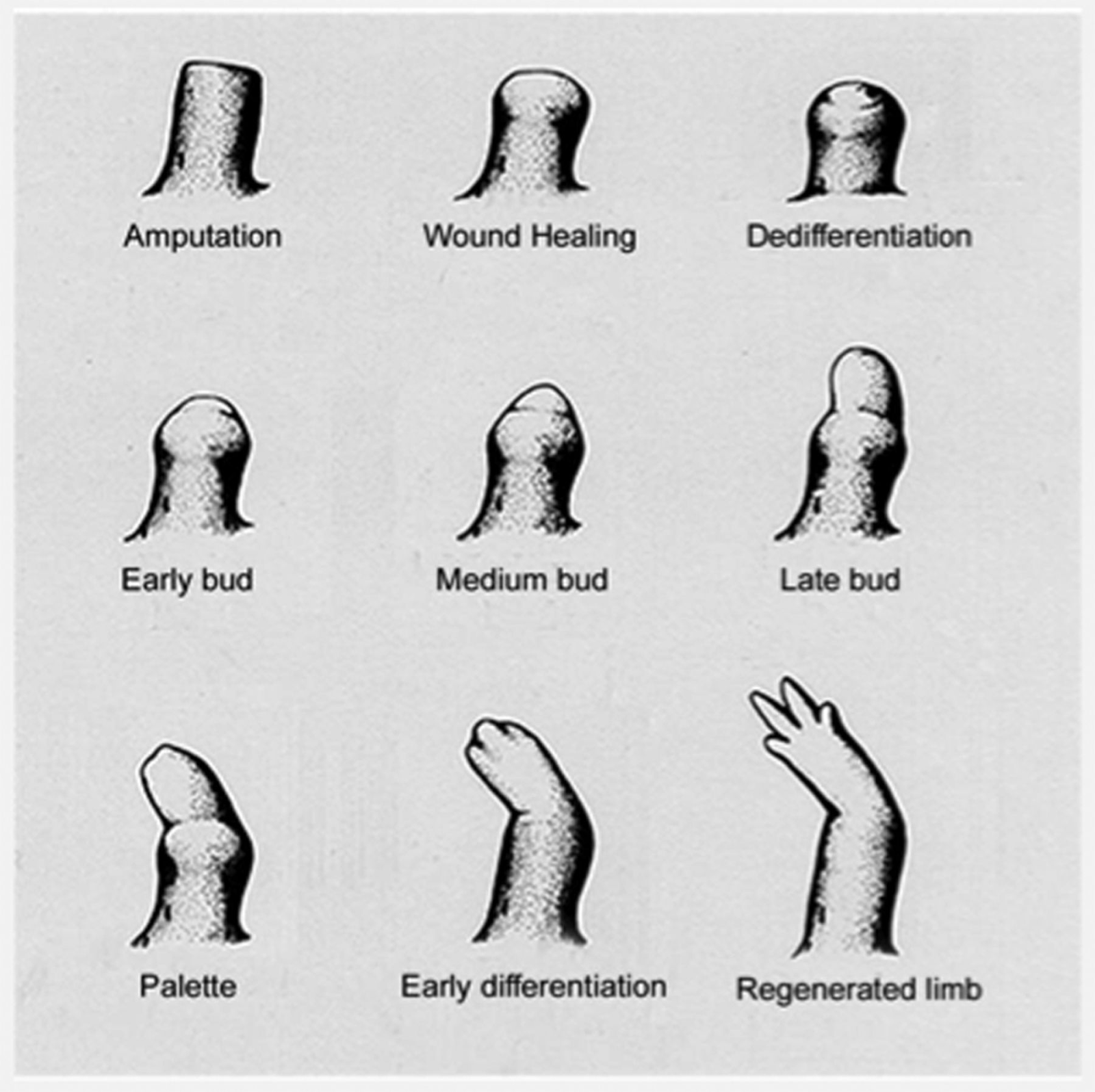
Figure 1. The stages of regeneration of a Newt following amputation (Iten & Bryant, 1973)
I. Apical Epidermis Cap & Blastema Formation
The process starts when one of its limbs are removed. Initially, the amphibians blood vessels tighten to minimize the amount of blood loss, and epidermal cells proliferate and cover the amputated area, similar to the idea of how a cut on a person may form a scab. An essential component called the apical epithelial cap forms from the initial epidermal cells, this occurs from the thickening of the epidermis (Brito, 2018). The apical epithelial cap functions as a signaling center which enhances blastema proliferation (Stocum, 2017). This is an important stage of the regeneration process because if this structure were to be damaged or missing then regeneration would seize to occur (Goss, 1969).
II. Limb Blastema Growth & Patterning
There are two mechanisms that explain how the limb blastema is formed since the origin is still up for debate. One source suggests that it accumulates through the activation of nearby stem cells (Simon & Tanaka, 2013). On the other hand, some say that the blastema is formed through the dedifferentiation of mature cells (Stocum, 2017). There are many different studies that explore which specific tissues contribute to the formation of the blastema, however, we will only focus on one of those in this section. A study reports that dermal fibroblast-derived cells contribute towards roughly 50% of the blastema makeup (Muneoka, Fox & Bryant, 1986).
Fibroblasts, which are a type of connective tissue, relocate to the middle of the injury and multiply until they become a blastema (Muneoka, Han & Garinder, 2008). This is caused by a protein called nAG (Kumar et al., 2007). Hox genes contained in the fibroblast cells determine which specific limb needs to be replaced based on its location in the body (Muneoka, Han & Garinder, 2008). The blastema proliferates creating numerous undifferentiated cells, which then become new blood vessels, bone, and muscle tissues (Kumar et al., 2007). The undifferentiated cells differentiate due to the Hox gene in the fibroblasts and become a new limb (Conger, 2008).
Macrophages are also a key component in limb regeneration. Macrophages are critical to the immune system and generate pro and anti-inflammatory signals. Studies have shown that reduced amounts of these cells in salamanders result in closure of the wound, but not limb regeneration. In a study, adding the macrophages back to the salamander resulted in regular limb regeneration (Godwin, Pinto & Rosenthal, 2013).
C. Hydra Description
Hydra, a bilayered freshwater polyp was subject to some of the first scientific investigations concerning regeneration in animals. Hydra belong to the Phylum Cnidaria, a phylum that includes corals, sea anemones, and jellyfish (Solomon, Berg, & Martin, 2002). Hydra has a tubular, radially symmetric body and exhibits distinct polarity along the oral/aboral axis (Zamaraev, 1956). The animal body is made up of two cell layers: the ectoderm and the endoderm - separated by an extracellular matrix; neurons and interstitial cells can be found between these two layers (King & Newmark, 2012). The cells of the Hydra belong to either the ectodermal, endodermal or interstitial cell lineage (Bosch, 2007). Scientists are especially interested in Hydra because of their remarkable regenerative abilities; tiny fragments of the animal tissue possess the ability to regenerate into complete organisms. Even dissociated Hydra tissue cells can reaggregate, reestablish polarity, and grow into whole new organisms (King & Newmark, 2012).
D. Mechanism - Morphallaxis
Regeneration in Hydra is accomplished through epithelial cells (Bosch, 2007). Additionally, a mesoglea which separates the two cell layers is required for the progression of regeneration. When a Hydra is cut in half, the head portion will regenerate a basal disc and vice versa. A Hydra can be spliced into many different pieces and both the head and basal disc components will regenerate accordingly. This is accomplished through polarity coordination via morphogenetic gradients.
In essence, there are signaling peptides that play an important role in the regeneration of the Hydra. Experiments have shown that there are two different types of signaling molecules; activators and inhibitors. These include head and foot activator peptides which enhance apical and basal regeneration respectively. There are different molecular pathways that can explain the biochemical mechanisms that describe Hydra regeneration, however, we will focus only on the MAPK pathway.
When the Hydras head is removed, the MAPK pathway becomes activated followed by the Wnt-beta-catenin pathway. This is all organized by the gastric tissue located in the regenerating tip of the Hydra. The MAPK pathway causes cell death in interstitial cells. This then causes Wnt genes to become activated in the progenitors of interstitial cells and carries over to the endodermal cells as well (Galliot, 2013). Resulting in organizing the cells involved with the head regeneration. Head regeneration in Hydra has shown to be flexible as decreases in cellular proliferation during Wnt activation did not have a significant effect on regeneration. Interstitial cycling and proliferation is important for this process. The cells located near the regenerating area increase proliferation and the cells farther away move closer. However, due to the flexibility of regeneration, the epithelial cells have been seen to regenerate the head without the interstitial cells, but this process takes longer (Galliot, 2013). It has been found that especially the HyWnt3 gene is important for head regeneration. Experiments that have silenced this gene have resulted in diminished head regeneration in the Hydra (Galliot, 2010). Investigators have taken an interest in these animal models as their remarkable regeneration capacities have inspired new ideas about how their regeneration can be applied to humans.
E. Human Application
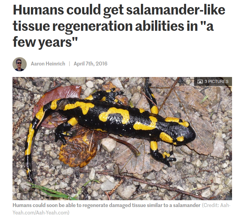 (Heinrich, 2016)
The regeneration in model organisms is applicable to humans as scientists can copy or use these ideas as a framework for future research. For example, using this idea of dedifferentiating cells, scientists are able to create induced multipotent stem cells out of fat cells. Through aspirating fat cells with two different solutions, the fat cells can convert back to a less specific state which can be used for wound healing. However, this cannot be used to regrow limbs, but this is a great step in the right direction for regenerative medicine. (Heinrich, 2016)
5. Regeneration in Humans
Regeneration in humans encompasses a wide range of clinical and research areas, from the use of stem cells to organ transplantation and therapeutic interventions. In humans, regeneration refers to the repair of a body structure, specifically tissues and organs, after a loss of function. During this process, stem cells can differentiate in order to replace lost tissue. They can also proliferate and transdifferentiate to assist in the regeneration process (Stark, 2018).
Current research in biomedicine focuses on animal regeneration and its application to humans. The vast majority of mammals, including humans, cannot regenerate whole tissue and complex organs. In contrast, other non-mammalian organisms can regenerate full organs and limbs, as shown above (Li et al., 2016). Research on model organisms can provide insight into the various processes involved in regeneration on a molecular level. What is currently known, however, is that some organs in the human body have some regenerative properties. Tissue regeneration is part of regular maintenance, where biochemical signals and stem cells create a regenerative environment for the tissue. However, this process is largely unknown. Other tissues have been shown to regenerate once stem cells are injected into the body. Biomedical research is currently focused in this area, as it can lead to the use of stem cell transplants and implanted materials for regeneration in humans (Stark, 2018).
Liver
The liver is the most well-known example of an organ with natural regenerative capabilities. Given the rising number of liver disease patients, regenerative mechanisms can be used to improve treatment given the cost, side effects, and lack of transplant options. The liver can regenerate from less than half of its original tissue because all liver cell types can proliferate, specifically hepatocytes and progenitor cells. When there is a large loss of liver tissue, rapid regenerative response will begin (Abdellatif & Shiha, 2017).
Mechanism - Compensatory
Hepatocytes are differentiated liver cells that have many metabolic functions. In response to tissue loss, existing hepatocytes enter the cell cycle under the influence of various growth factors such as AP-1 and STAT-3 (Fabrikant, 1968). Hepatocyte proliferation is initiated in the outer region of the liver and then moves towards the center (Abdellatif & Shiha, 2017). After removing two-thirds of the pancreas in rodents, it has been found that hepatocytes undergo 1.4 rounds of replication on average to restore normal liver size, taking 5-7 days. In humans, this process takes 3-4 weeks (Michalopolous, 2007).
The liver also has hepatic progenitor cells (HPCs) that can be activated during chronic liver damage. The HPCs are can fully differentiate into hepatocytes and bile duct cells. Other studies have demonstrated that these cells can also transdifferentiate into hepatocytes when they are unable to proliferate during an injury (Evarts et al., 1996).
Spinal Cord
The spinal cord is an example of induced regeneration and does not occur naturally in the human body. The aim of spinal cord regeneration is to reconnect damaged nerves and reverse paralysis. Currently, there is no cure for spinal cord injuries. This is because the nerves become blocked by severe scar tissue that does not allow a natural molecular regeneration process (Kabu et al., 2015). Research conducted by Li et al., (2017) has shown promising results, using synthetic and mesenchymal stem cells for scar tissue removal. However, these cells rapidly degrade and cannot withstand high temperatures.
One of the most widely researched therapies is the use of olfactory ensheathing cells (OECs), which are cells involved in axon extension and promote receptor signalling for the human sense of smell. They have been found to regulate neuroinflammation and promote axon growth. Thus, they have been used in animal transplant studies as a treatment for spinal cord injury (Chehrehasa et al., 2010). Though many studies have reported neurological benefits, the research is inconsistent. Furthermore, there are many populations of OECs and purification methods must be put in place (Deumans et al., 2018).
6. Medical Applicants / Treatments
Regenerative medicine is an ongoing interdisciplinary field of research that aims to develop treatments that replace- or restore organ functions and tissue damages from the effects of diseases, injuries, aging, or congenital defects [23]. Since the early 1950s, regenerative medicine has been studied and practiced within the health and science industries in hopes to fully restore the functions of human tissues and organs. The first cell transplantation: bone marrow transplantation, and the first organ transplantation: kidney transplantation, has sparked further insight and discovery within this field of medicine [25]. With the rising demand for healthy tissues and organs, many regenerative medicine therapies have found its way into clinical settings and commercial usage.
a. Current Commercial Usage
An increased amount of regenerative medicine therapies has received either clearance or approval from the FDA- Food and Drug Administration (Table 1). Carticel is one of the earlier products that made its way into commercial usage. To treat defects within the focal articular cartilage, Carticel uses autologous, from the same individual, chondrocytes. Essentially, cells that produce cartilage in the knees are applied to the body to combat the chondral defect [20]. Apligraf serves as a living, bi-layered substitute for human skin [2]. Apligraf uses allogeneic, genetically similar but not identical, stem cell transplantation, a treatment where stem cells from a healthy individual is applied to the damaged skin of the recipient [18]. Similarly, Dermagraft uses human cells, fibroblasts, to treat damaged cells or skin tissue caused by foot ulcers [24]. Celution is a machine that efficiently produces adipose-derived regenerative cells (ADRCs) which are aimed to treat wounds or assist in wound healing [8]. Both Regranex and Infuse, infuse bone graft, and inductors use growth factors that allow for the regeneration into biomaterials [18]. Regranex specifically uses platelet derived growth factors while Infuse, infuse bone graft, and inductors use bone morphogenetic protein 2- growth factors [18]. Though these products have been approved by the FDA and have shown evidence for assisting in healing and regeneration, they still fail to fully resolve the issue, whether it be an injury, disease, or congenital defect. Complications and critiques have also came about concerning some of these treatments. Thus, research in regenerative medicine therapies remains on-going with the aim to improve upon current strategies and develop more reliable and efficient treatments.

b. Addressing Limitations and Steps Towards Complete Human Organ or Tissue Recovery
Regenerative medicine continues to develop as there are variety of limitations that hinder the full treatment of non-functional tissues and organs. For example, the behavior of stems cells needs to be strictly controlled for in order to improve safety and efficacy post-transplantation treatments [18]. To do this, researchers are looking into building microenvironments that include specific cues that allow for direct manipulation of target cells [18]. One of the biggest limitations hindering the complete treatment of dysfunctional or damaged tissues and organs is immune reactions, specifically the rejection of regenerative treatments from immune responses [18]. Further knowledge and expertise in the human immune system, along with a clearer understanding of how age, disease, and one’s biological environment affect regeneration is needed to enhance existing regenerative treatments. As development continues, our health care system may be able to provide tissue-engineered heart muscles for damaged heart tissue, infuse cells into a patient’s existing matrix to improve lost functionality, regenerate insulin-producing pancreatic islets to assist with the effects of diabetes, and many more [23].
References
[1] Abdellatif, H., & Shiha, G. (2017). Liver Regeneration: Summary of Involved Cell Types. Journal of Stem Cell and Regenerative Biology, 3(1), 0-0.
[2] Apligraf. (2010). What is Apligraf? Retrieved from http://www.apligraf.com/professional/what_is_apligraf/index.html
[3] Bosch, T. C. (2007). Why polyps regenerate and we don't: towards a cellular and molecular framework for Hydra regeneration. Developmental biology, 303(2), 421-433.
[4] Cai, S. A., Fu, X., & Sheng, Z. (2007). Dedifferentiation: a new approach in stem cell research. AIBS Bulletin, 57(8), 655-662.
[5] Chehrehasa, F., Windus, L. C., Ekberg, J. A., Scott, S. E., Amaya, D., Mackay-Sim, A., & St John, J. A. (2010). Olfactory glia enhance neonatal axon regeneration. Molecular and Cellular Neuroscience, 45(3), 277-288.
[6] Deumens, R., Van Gorp, S. F. J., Bozkurt, A., Beckmann, C., Führmann, T., Montzka, K., … & Brook, G. A. (2013). Motor outcome and allodynia are largely unaffected by novel olfactory ensheathing cell grafts to repair low-thoracic lesion gaps in the adult rat spinal cord. Behavioural brain research, 237, 185-189.
[7] Evarts, R. P., Hu, Z., Omori, N., Omori, M., Marsden, E. R., & Thorgeirsson, S. S. (1996). Precursor-product relationship between oval cells and hepatocytes: comparison between tritiated thymidine and bromodeoxyuridine as tracers. Carcinogenesis, 17(10), 2143-2151.
[8] Fraser, J. K., Kicok, K. C., Shanahan, R., Zhu, M., Miller, S., & Arm, D. M. (2014). The Celution System: Automated Processing of Adipose-Derived Regenerative Cells in a Functionally Closed System. Adv Wound Care, 3(1), 38-45.
[9] Galliot, Brigitte(Nov 2013) Regeneration in Hydra. In: eLS. John Wiley & Sons Ltd, Chichester. Retrieved from http://www.els.net [doi: 10.1002/9780470015902.a0001096.pub3]
[10] Godwin, J. (2014). The promise of perfect adult tissue repair and regeneration in mammals: learning from regenerative amphibians and fish. BioEssays, 36(9), 861-871.
[11] Godwin, J. W., Pinto, A. R., & Rosenthal, N. A. (2013). Macrophages are required for adult salamander limb regeneration. Proceedings of the National Academy of Sciences, 110(23), 9415-9420.
[12] Gilbert, Scott F.; Barresi, Michael J. F. (2016). Developmental Biology (11th ed.), 701–702.
[13] Heinrich, A. (2016, April 08). Humans could get salamander-like tissue regeneration abilities in “a few years”. Retrieved from https://newatlas.com/reprogramming-stem-cells/42697/
[14] King, R. S., & Newmark, P. A. (2012). The cell biology of regeneration. J Cell Biol, 196(5), 553-562.
[15] Kragl, M., Knapp, D., Nacu, E., Khattak, S., Maden, M., Epperlein, H. H., & Tanaka, E. M. (2009). Cells keep a memory of their tissue origin during axolotl limb regeneration. Nature, 460(7251), 60.
[16] Li, J., Zhang, S., & Amaya, E. (2016). The cellular and molecular mechanisms of tissue repair and regeneration as revealed by studies in Xenopus. Regeneration, 3(4), 198-208.
[17] Lu, J., Féron, F., Mackay‐Sim, A., & Waite, P. M. (2002). Olfactory ensheathing cells promote locomotor recovery after delayed transplantation into transected spinal cord. Brain, 125(1), 14-21.
[18] Mao, A. S., & Mooney, D. J. (2015). Regenerative medicine: Current therapies and future directions. Proc Natl Acad Sci U.S.A., 112(47), 14452-14459. Retrieved from https://www.ncbi.nlm.nih.gov/pmc/articles/PMC4664309/
[19] Metcalfe, A., & Waugh, N. (2017). Autologous chondrocyte implantation in the knee: systematic review and economic evaluation. Health Technology Assessment, 21.6.
[20] Mistry, H., Connock, M., Pink, J., Shyangdan, D., Clar, C., Royle, P., Court, R., Biant, L. C., Michalopoulos, G. K. (2007). Liver regeneration. Journal of cellular physiology, 213(2), 286-300.
[21] Morrison, J. I., Lööf, S., He, P., & Simon, A. (2006). Salamander limb regeneration involves the activation of a multipotent skeletal muscle satellite cell population. The Journal of cell biology, 172(3), 433-440.
[22] Muneoka, Ken; Han, Manjong; and Garinder, David M. “Regrowing Human Limbs.” Scientific American. April 2008. (Oct. 28, 2008). Retrieved from http://www.sciam.com/article.cfm?id=regrowing-human-limbs
[23] NIH. (2018). Regenerative Medicine. Retrieved from https://report.nih.gov/nihfactsheets/ViewFactSheet.aspx?csid=62
[24] Organogensis inc. (2013). Treating Your Diabetic Foot Ulcer. Retrieved from http://www.dermagraft.com/patient/about-dermagraft/
[25] Sampona, G., Guraya, S. Y., Forgione, A. (2015). Regenerative medicine: Historical roots and potential strategies in modern medicine. Journal of Microscopy and Ultrastructure, 3(3), 101-107.
[26] Singh, V. K., Saini, A., Kalsan, M., Kumar, N., & Chandra, R. (2016). Describing the stem cell potency: the various methods of functional assessment and in silico diagnostics. Frontiers in cell and developmental biology, 4, 134.
[27] Stark, J. F. (2018). Perspectives on human regeneration. Palgrave communications, 4(1), 66.
[28] Slack, J. M. (2007). Metaplasia and transdifferentiation: from pure biology to the clinic. Nature reviews Molecular cell biology, 8(5), 369.
[29] Solomon, E., Berg, l., Martin, D. (2002) Biology 6th edition. Brooks/Cole Publishing.
[30] Zamaraev, V. N. (1956). Distortion of polarity in hydra. Bulletin of Experimental Biology and Medicine, 42(6), 1051-1053.